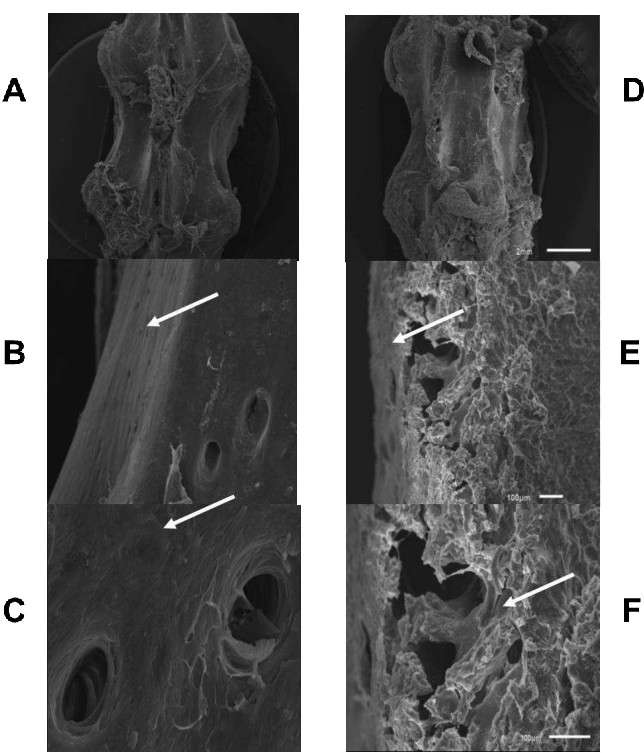
Fig. 3. Evaluation of the vertebral bone ultrastructure through Scanning Electron Microscopy (SEM). SEM was used to analyzed whether there were a change into bone vertebrae bone ultrastructure trying to conciliate the data we obtained so far (all bones use from each, 3 and fourth verterbral bone, animal were cutted three consecutive times and several pictures were taken form than. We consider n=3 the number of animals not the number of images taken). From A to C we observed representative figures taken from MOCK Treated rat (n=3) on of 2 mm (A), 100 µm, and10 µm, respectively. From D to F it is observed images taken from vertebrae of TBT-treated animals (also n=3) at same order of magnificence described previously. Note deterioration of bone matrix and decreased compact bones in TBT-treated animals when compared to controls (arrows) is revealed at level of mm. When examined at µm scale inner bone (spongy bone) shows complete deterioration. Another thing to be pointed out is that external layer (compact bone) is clearly diminished, strongly suggesting that bone in affected deeply by TBT.